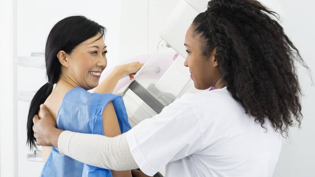
Breast cancer screening is moving away from one-size-fits-all guidelines toward personalized plans based on individual risk factors. While general recommendations suggest women begin screening around age 40, healthcare professionals now evaluate each patient in context. This approach considers personal and family history, breast density, and other specific risk factors to determine when and how someone should be screened, including whether newer options are available for those at higher risk.
Building a Personal Risk Profile
Finding out about family history is a key step in the screening process. During routine visits, healthcare teams ask about relatives who have had breast cancer, specifically first-degree relatives like mothers or sisters. This information can lead to a referral for a genetic counselor, who reviews family history and completes a risk assessment. Genetic testing may then be offered based on that analysis. Open conversations with family members about diagnoses, cancer types, and treatments can provide valuable details for the healthcare team.
Genetics only accounts for a small portion of breast cancer cases, however. Inherited factors contribute to just 5% to 10% of diagnoses. Other factors considered include the age a person started menstruating and the age menopause began. When these factors are combined with family history, they provide a more complete picture of a patient’s risk profile.
While a dense family history might suggest a need for closer monitoring, the presence of dense breasts alone does not automatically require supplemental imaging. The decision depends on how density interacts with other risk factors. For some individuals, particularly those at higher risk, screening may include breast MRI, molecular breast imaging, or contrast-enhanced mammography.
How Technology Is Changing Detection
Advances in imaging technology are improving the evaluation of breast tissue. Three-dimensional mammography, or digital tomosynthesis, adds a third dimension to traditional two-dimensional scans. This allows radiologists to examine tissue in greater detail and spot findings like calcifications or areas that are difficult to see. Artificial intelligence is also being used to model and detect tissue patterns, helping professionals evaluate changes over time.
These technological improvements may help reduce unnecessary procedures. In the past, a questionable area on a standard mammogram often led to a biopsy or lumpectomy. With better detection capabilities, some findings can now be monitored with follow-up imaging to determine if they remain stable rather than undergoing immediate invasive testing.
Because every patient’s risk profile is unique, screening plans differ significantly from person to person. A friend might be advised to have additional imaging due to specific risk factors, but that does not mean the same approach is appropriate for everyone. Healthcare teams combine age, family history, breast density, and other factors to determine the most suitable screening strategy for each individual.